Monthly Archives

January 2014

‘Beatrix’ Morris-Behind The Scenes Of Her Dental Procedure!

By Our Blog No Comments

As a precursor to Dental Awareness month in February, Dr. Morris thought it would be interesting for you to see behind the scenes of his dog Beatrix’ dental procedure. I will caution you, some people may find a few of these images a bit graphic but we think it’s a great opportunity to see what happens during a dental procedure.

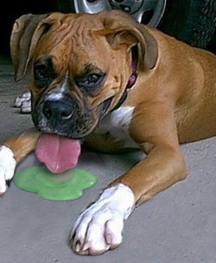
A little background: Beatrix is an 8 year old Jack Russel. For those of you who haven’t met Bea, she is a 3 legged wonder dog!

In December Dr. Morris found a small lump under her left eye. After a few days this lump went away, but it quickly returned! Dr. Morris suspected she may have a tooth root abscess and she was scheduled for a dental procedure. Here is what happened:

B in cage


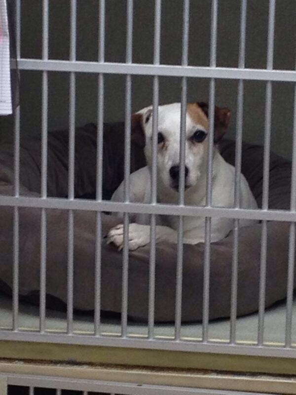
The night before her procedure Bea came in for some pre-op testing. She had blood and urine profiles done to make sure she was healthy for the anesthetic. Then Bea stayed over with us! She was very cozy in her bed. so cute!


B pre-op


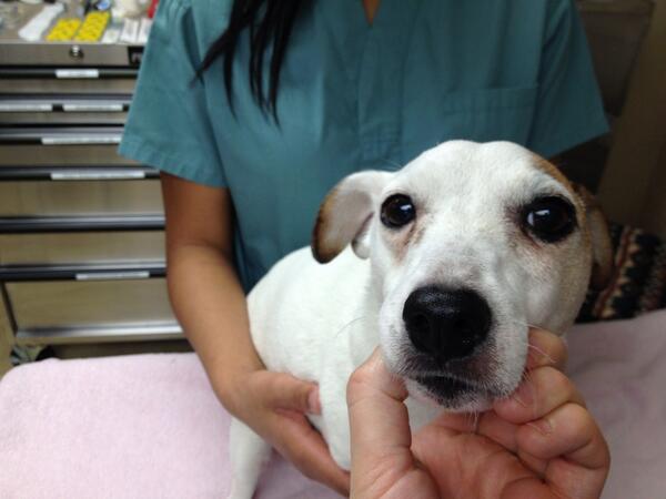
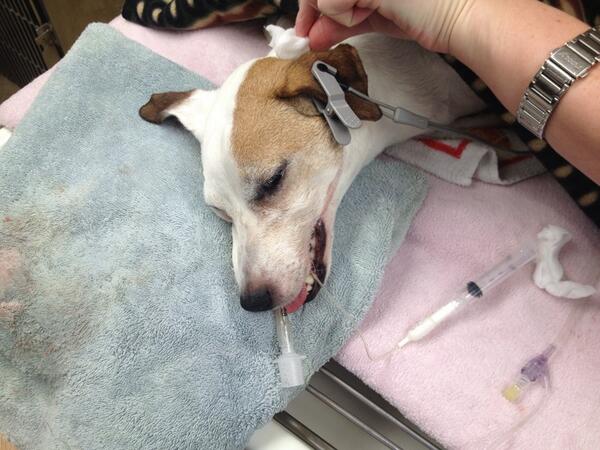
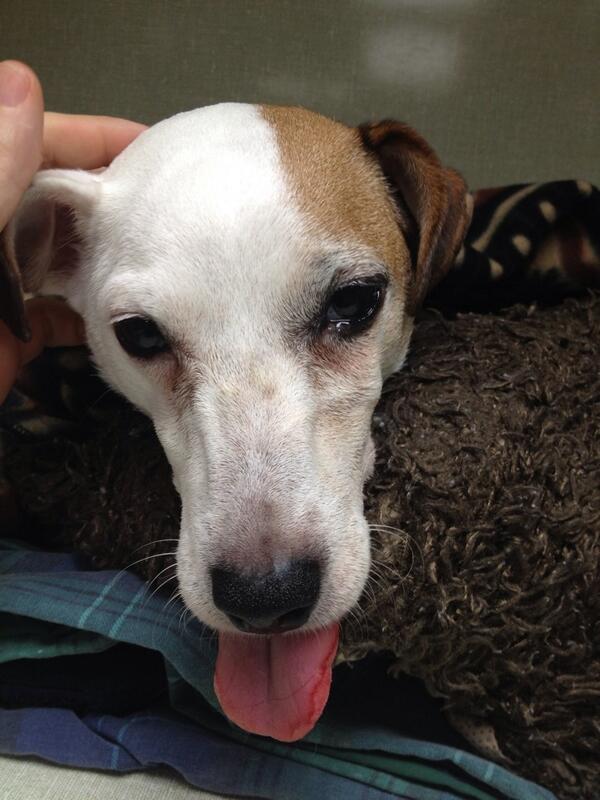
In the morning Dr. Morris gave her another full physical exam. This is Bea on the exam table in our treatment room. If you look closely you can see the lump under her left eye. It’s at this time when the Registered Technician gives Bea an injection of a sedative and pain control to calm her before they start.


B sedated


Here is Bea now sedated. You can see how sleepy she is. The Technician has placed her IV catheter and fluids and she is ready to go under anesthetic. It is also at this time where the technician and assistant will attach some of her monitoring equipment. We us a doppler machine that allows us to hear her heart beat at all times. We also use the doppler to closely monitor her blood pressure during the procedure.

Once her doppler is on, the technician will administer an injectable anesthetic into her IV. This will sedate her enough to allow the technician to place a breathing tube in her airway and attach the gas anesthetic.


B ready for rads

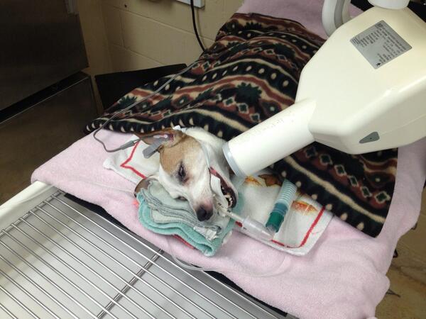
Bea is now under full general anesthetic. At this time, more monitoring equipment is placed on her. You can see a grey clip on her ear; this is a pulse ox monitor and it monitors the amount of oxygen in her blood as well as her pulse.  Bea is quite snugly under all those blankets! When animals are put under general anesthetic their temperatures will drop. We do our best to prevent this by using warming devices and lots of blankets. The technician will now take x-rays of all of her teeth.


B first rad


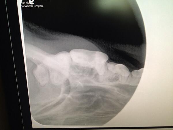
Here is the first x-ray of that questioned tooth. She has chipped the crown of the tooth but in this angle the root is missing?


B second rad

Here is a new angle! There’s the missing root. The dark shadow around the tip of the root is due to bone loss and infection!


B third rad


Here is the other half of the problem tooth. More signs of infection around these root tips as well. The best treatment is to extract the tooth.


B nerve block


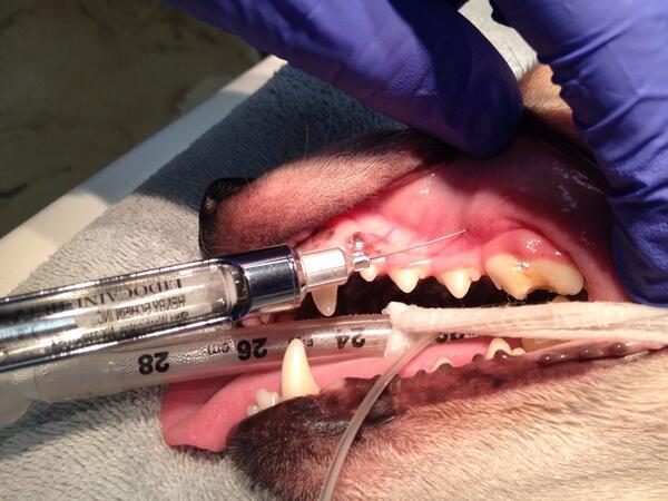
After the technician cleans and polishes all of her teeth, making sure she cleans under the gum line-the exact same as when you and I go to the dentist. Dr. Morris freezes the nerves around the tooth. This will make the extraction pain free. Freezing the tooth allows Dr.Morris to extract the tooth without having to deepen her anesthetic, making the procedure as safe as possible.


B post extraction


After 40 minutes of extracting, the tooth is finally out and Dr. Morris places stitches. These stitches will dissolve on their own in a few weeks.


B waking up

The procedure is over and her gas anesthetic is turned off. All of her monitoring equipment will stay on until she is completely awake. Dr. Morris, the technician and the assistant are sitting with her as she wakes up. Bea slowly wags her tail, she can hear Dr.Morris’ voice as she is coming around.

Remember that her mouth is still frozen and we gave her an injection of pain control before her procedure started. This will keep her very comfortable during her recovery. An injection of a long lasting pain medication is given to her right at recovery and Dr.Morris will also continue to give her some mild pain control while she is at home.


B recovery


Bea is now back in her recovery cage. Still a little groggy, but very comfortable. The technicians and assistants will continue to keep a close eye on her. Once is is fully awake the assistant will take her out for a walk and will offer her some food. She will be eating soft food only for a couple of weeks to allow her extraction site to heal.


photo (11)


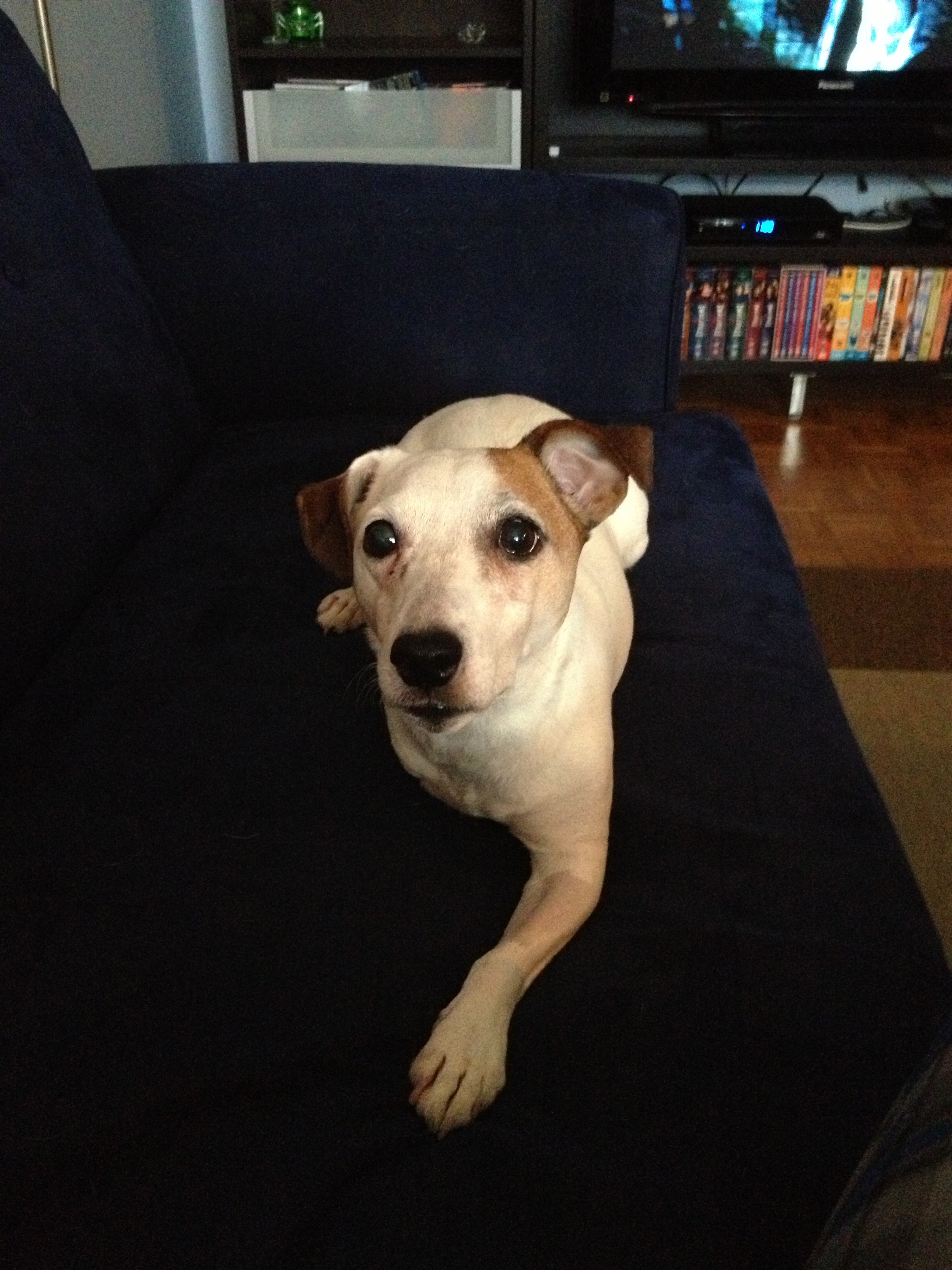
It’s been a couple of days since her dental procedure and Bea is back at home and feeling better than ever! Look how happy she is!!


Dental care is one of the best ways we can help ensure our pets live a long and healthy life. If left untreated, the infection in her mouth would have effected Bea’s internal organs and continued to cause her pain. Take a look in your pets’ mouth today! some signs of dental disease are:

  • Yellow/brown tartar build up
  • red or bleeding gums
  • foul smelling breath
  • missing or wiggly teeth
  • lumps under the eyes
  • difficulty eating or pawing at their mouths

If you are concerned about your pets’ oral care, please see your veterinarian.

We would like to thank Beatrix Morris for allowing us to document her procedure. 


Antifreeze Poisoning…What You Need To Know.

By Our Blog No Comments

This time of year most of us in the Bloor West Village are forced out in the freezing weather to warm up and de-ice our cars before our trip to work. Many times this ritual includes topping up our windshield washer fluid and antifreeze. This task, if not done safely can have deadly consequences for our pets and wildlife. Antifreeze contains ethylene glycol. It is a sweet tasting, odourless liquid and is highly toxic to animals.

Ethylene glycol can also be found, in lower concentrations, in some windshield de-icing agents, hydraulic brake fluid, motor oils, solvents, paints, film processing solutions, wood stains, inks, printer cartridges, etc.

Unfortunately animals are attracted to the ethylene glycol by its sweet taste and will drink it voluntarily if they have access. Ethylene glycol is so toxic that only a tiny amount can have deadly consequences if ingested.  As little as 1 tablespoon can be fatal to a medium sized dog!

What are the signs of ethylene glycol poisoning?

Early signs of ethylene glycol poisoning are called “Stage 1”, and can be seen within 30 minutes of toxin ingestion. The signs include:

  • lethargy
  • vomiting
  • in-coordination (walking drunk)
  • excessive urination
  • excessive thirst
  • hypothermia (low body temperature)
  • seizures
  • and coma.

In “Stage 2” (which occurs 12 to 24 hours after ingestion), some of the signs seem to dramatically improve, luring pet owners into a false sense of security. However, during this stage, pets become dehydrated, and develop an elevated breathing and heart rate.

“Stage 3” occurs 12-24 hours after ingestion in cats, and 36-72 hours after ingestion in dogs. At this stage:

  • signs of severe kidney dysfunction, which is characterized by swollen, painful kidneys and the production of minimal to no urine, may occur.
  • Progressive depression
  • lethargy
  • lack of appetite
  • vomiting
  • seizures
  • coma
  • death 

It is critical that you bring your pet to a veterinary clinic if you know or even suspect that they have consumed ethylene glycol, or if they are exhibiting any of the early symptoms. Do not wait; time is of the essence and immediate treatment is essential! the antidote only has a narrow time period to work. Left untreated, the animal will die.

Keep your pets and your local wildlife safe by storing your antifreeze where they can not access it, clean up any spills immediately and leave all pets in the house when topping up your reservoirs.

Let’s keep ourselves warm and our pets safe and let the countdown to spring begin!


Written by Ashley Docherty, RVT and Practice Manager at Bloor Animal Hospital.  This blog is based on material written by: Kristiina Ruotsalo, DVM, DVSc, Dip ACVP & Margo S. Tant BSc, DVM, DVSc & Justine A. Lee, DVM, DACVECC

© Copyright 2012 Lifelearn Inc. Used and/or modified with permission under license.


Your Pets’ Weight…a Heavy Subject

By Our Blog No Comments

In the new year, some of us make resolutions for ourselves. But what about our pets? Now is a good time to have your pets weight evaluated and work on trimming down for the spring.

Your pets’ weight is one of the most important things we can monitor throughout our pets lives. Maintaining our pet’s ideal weight is a simple and inexpensive ways we can prevent many health problems in life. It’s a way we can ensure our pets live a comfortable, and long life.

Today veterinarians are finding a large increase in overweight or obese pets, more now then ever. Weight gain is usually associated with an increase of calories consumed, combined with a lack of exercise. A few extra pounds gained for a person may not be very significant; however, for pets, weight gain can mean major health risks and even a shorter life span.

Obesity can cause the same health problems in pets as it does in people such as: diabetes, joint problems, lower urinary tract disease in felines, strain on kidneys, heart and liver, and they can be at a higher risk of complications during procedures which require anesthetic.

A question you may ask is, “How do I know if my pet is becoming over weight?”

You should be able to feel their ribs under a thin layer of fat, but not be able to see them. They should have an hourglass figure when viewed from above, and their abdomen should be tucked up under their rib cage.

 If you believe your pet is, or is at risk of becoming overweight, the most important thing to do is to book an appointment with your veterinarian. There could be a medical reason for your pets’ weight gain and this must be ruled out before starting your pet on a diet. It is very important to never start your pet on a diet before consulting with your veterinarian. We need to change their eating and exercising habits gradually.

A calculation is used to determine the appropriate amount of calories your pet needs to consume in a day. A diet is chosen for your pet based on the amount of weight your pet needs to loose, whether or not they like the food, and whether it works with your pet’s metabolism. Your pets’ weight needs to be monitored closely by weighing them monthly. Once your pet has reached his or her weight loss goal, we can then gradually change them to a weight maintenance diet.

 Remember, an overweight pet is not a healthy pet. They don’t gain the weight overnight; therefore we can’t expect them to loose the weight overnight. Together with the help of your veterinarian, patience and perseverance you can achieve your pet’s weight loss goal.

Written by Ashley Docherty Registered Veterinary Technician & Practice Manager at the Bloor Animal Hospital

2387 Bloor St West